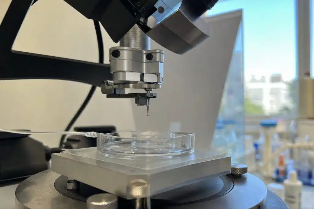
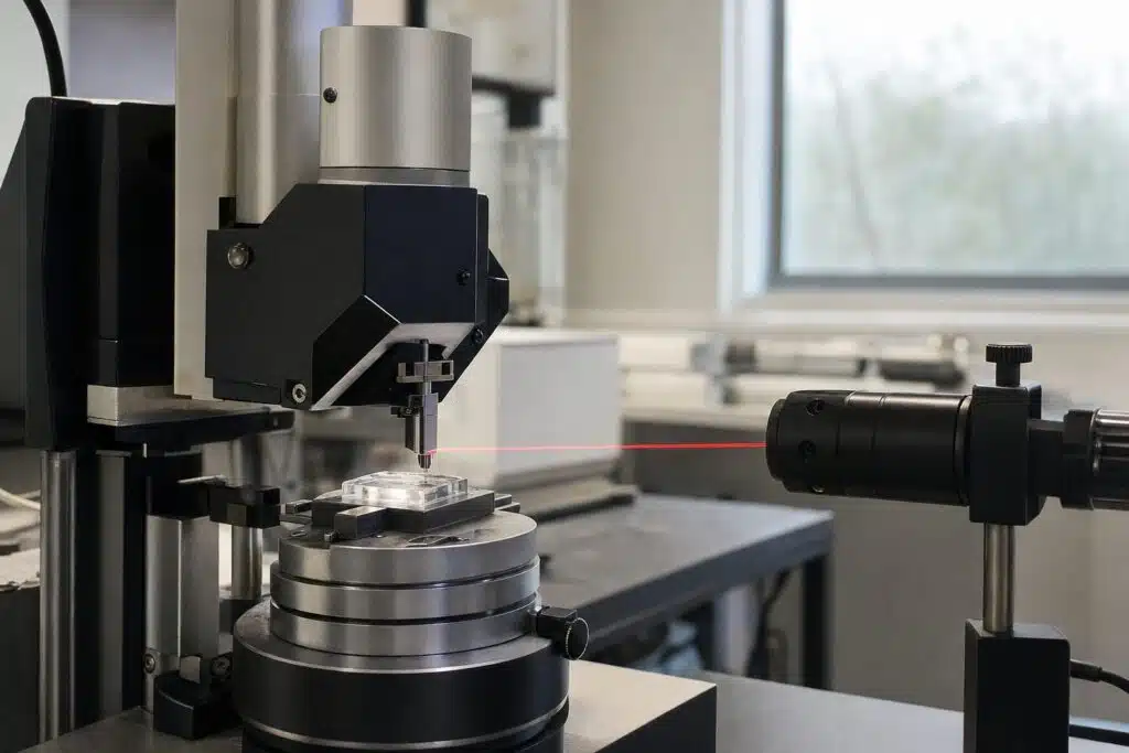
An atomic force microscope (AFM) works by scanning a very sharp tip, mounted on a flexible cantilever, across a surface and using a laser to read how the cantilever bends, which builds a three-dimensional map of the surface at nanometre scale. Unlike an optical or electron microscope, the AFM does not form an image from light or electrons. It feels the surface, much as a record player stylus follows a groove, and that is why it works in air, in liquid and on insulating samples. This guide explains each part, the feedback loop, the main imaging modes and what resolution to expect. For the full set of modes, see AFM imaging modes explained.
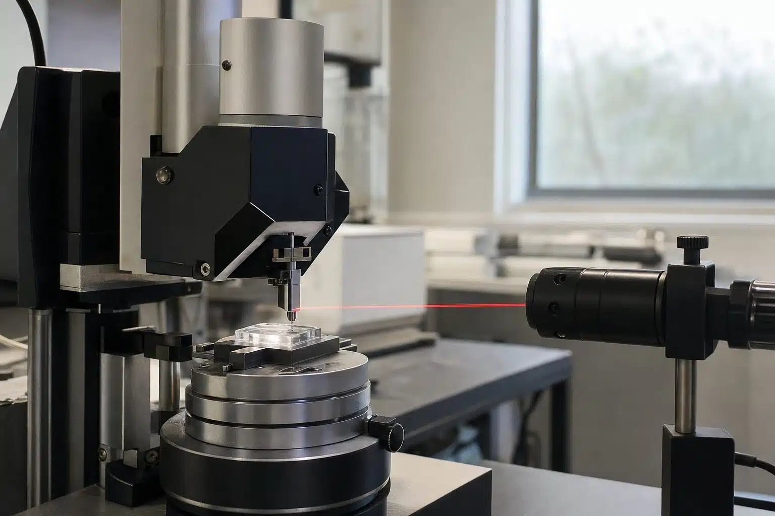
Table of Contents
What Are the Main Parts of an AFM?
- Cantilever and tip. A microfabricated cantilever, typically silicon or silicon nitride, carries a tip whose apex can be a few nanometres wide. See our AFM cantilever guide for how to choose one.
- Laser and photodiode. A laser reflects off the back of the cantilever onto a position-sensitive photodiode, which reads tiny bends.
- Piezo scanner. Piezoelectric ceramics move the tip or sample in X, Y and Z with sub-nanometre steps.
- Controller and feedback loop. Electronics keep the tip-sample interaction constant by adjusting Z.
- Vibration isolation. A table or enclosure shields the head from floor vibration and air flow.
The AFM technology page shows how these elements combine in research systems.
How Does the Feedback Loop Build an Image?
The tip approaches the surface until forces between tip and sample bend the cantilever slightly. The controller then sets a target for that bend, called the setpoint. As the scanner moves sideways, the surface height changes, the bend departs from the setpoint and the feedback loop moves the scanner up or down to restore it. The recorded Z movement at every point becomes the height image. The Wikipedia article on atomic force microscopy gives the physics of the tip-sample forces in more detail.
What forces act on the tip
At close range the tip feels repulsion from overlapping electron clouds. At slightly larger distances it feels attractive van der Waals forces, and in air a thin water layer adds capillary force. Different modes make use of different parts of this force curve.
What Imaging Modes Are Common?
| Mode | How the tip behaves | Good for |
|---|---|---|
| Contact mode | Stays in contact, cantilever bend is held constant | Hard, flat samples and friction maps |
| Tapping (dynamic) mode | Oscillates and taps briefly at each point | Soft samples, polymers, biology |
| Phase imaging | Records the phase lag of the oscillation | Material contrast on mixed surfaces |
| Force spectroscopy | Pushes and retracts at one spot | Stiffness and adhesion |
| Electrical modes | Measure conductivity or surface potential | Semiconductors and batteries |
Dynamic modes cut lateral forces, so they are gentler on soft samples. Electrical variants include conductive AFM and Kelvin probe force microscopy, described in our Kelvin probe guide.
What Resolution Can an AFM Reach?
Vertical resolution is often below a nanometre, since the scanner and sensors can resolve tiny height changes. Lateral resolution depends on the tip radius and the sample, typically a few nanometres, and atomic resolution is possible on flat crystals under good conditions. Our article on AFM resolution explains the limits. Because the tip is a physical shape, a blunt tip broadens narrow features, which is called tip convolution.
What Do Research AFM Systems Offer?
Nanosurf describes the CoreAFM as an integrated unit with a flexure-guided scanner, an XYZ sample stage, a camera for sample observation, an active vibration table and airflow shielding, plus a 24-bit controller. Standard modes include static force, dynamic force, lateral force, phase imaging, liquid imaging, force modulation, MFM, spectroscopy and lithography. The SmartSPM from the same catalog is aimed at higher performance: it uses a single 100 micrometre scanner for large scans and molecular or atomic resolution, and fully automated laser to photodiode alignment, according to the manufacturer. If budget is the question, our overview of atomic force microscope prices explains what drives cost.
Where Is AFM Used?
Materials scientists use AFM to measure roughness of coatings, thin films and polished surfaces, and to see grain structure and defects. Polymer researchers map stiffness and phase to separate components in a blend. Battery and semiconductor labs use electrical modes to find conductive paths and surface potential differences. In life science, AFM images molecules, membranes and cells in liquid, and force curves measure how strongly a ligand binds to a surface or how stiff a cell is.
Sample preparation basics
Samples must be firmly fixed, because a loose particle will move under the tip. Flat substrates such as mica, silicon or glass give the cleanest background for particles and molecules. Rough samples with steep features need slower scans and longer tips, and very soft samples benefit from low setpoints and dynamic modes.
Reporting data
Always state the scan size, scan rate, mode, probe type and any image processing. Roughness values depend on the scan size, so compare them only when the scan areas match.
How Is AFM Different From STM and SEM?
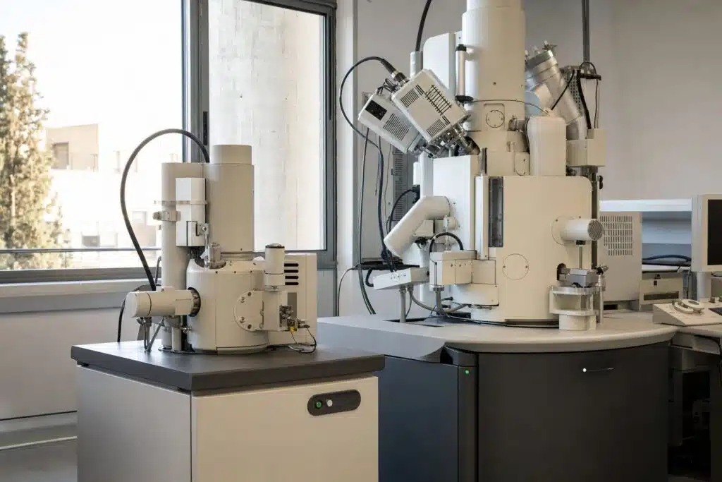
AFM works on insulators as well as conductors, while the scanning tunneling microscope needs conductive samples, a trade-off covered in AFM vs. STM. An electron microscope gives fast, wide-field views and elemental data but needs vacuum, while an AFM gives true height data and works in liquid.
How Do You Get a Good Image?
Choose the tip for the job: stiff and sharp for hard samples, softer cantilevers for delicate ones. Start with a gentle setpoint, check that the trace and retrace lines match, and reduce the scan speed if they do not. Level the image by plane fitting after scanning, but avoid aggressive filtering that can create features. Replace the tip when images show repeated shapes or lose sharpness.
Common artefacts
Double tips show repeated copies of features. Feedback oscillation shows ringing at edges. Thermal drift stretches images, so let the system settle before scanning. Contamination on the tip creates streaks and sudden changes in contrast.
Looking for an AFM for research or teaching?
Frequently Asked Questions
How does an atomic force microscope work?
A sharp tip on a cantilever scans the surface while a laser and photodiode measure the cantilever bend. A feedback loop keeps the interaction constant, and the scanner movement forms the height image.
What can an AFM measure?
It measures surface topography in three dimensions and, in other modes, stiffness, adhesion, friction, conductivity and surface potential.
Can an AFM image in liquid?
Yes. Many AFMs operate in liquid, which makes them useful for biological samples and electrochemistry.
What is the resolution of an AFM?
Vertical resolution is often below a nanometre, and lateral resolution is typically a few nanometres, set mainly by the tip radius.
What is tapping mode?
The cantilever oscillates and touches the surface only briefly at each point. This reduces lateral forces and suits soft samples.
Does an AFM need a vacuum?
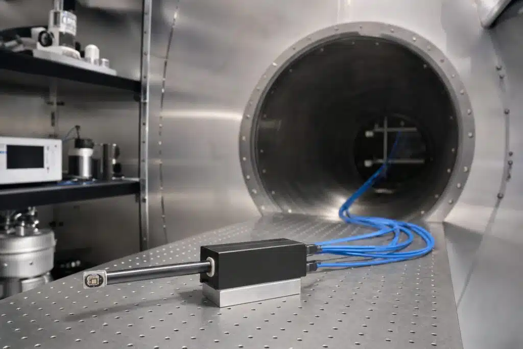
No. It works in air, liquid or vacuum, which is a main advantage over electron microscopy.
Last updated: November 2026